Scientists grow human eye parts to determine how we see in color
Lab-grown organoids reveal how the eyes develop and may lead to therapies for diseases such as color blindness.


Eye can barely believe it, but it's true.
Researchers at John Hopkins University in Maryland created eyeball parts from stem cells in the hopes of better understanding the how and why we developed "trichromatic vision" -- the ability to see in red, blue and green. The study was published in Science on Oct. 12.
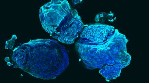
Organoids are built in vitro from a small number of stem cells in a 3D suspension, which eventually multiply to form something akin to an organ system.
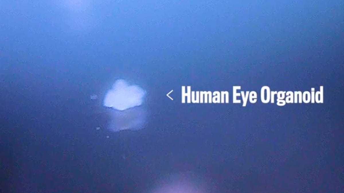
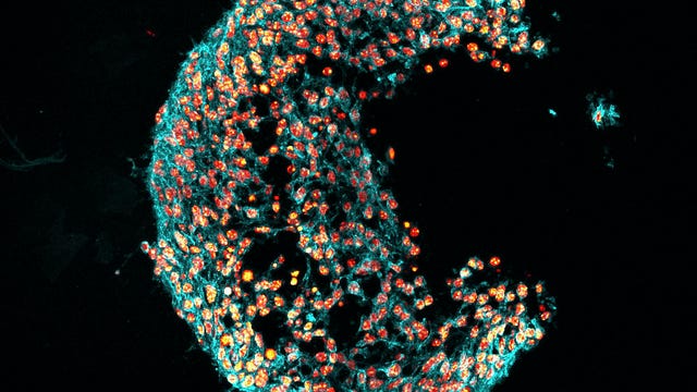
The eye organoid used in the John Hopkins study produces a miniature retina, the layer of cells at the back of the eyeball that process light, creating the electrical impulses the brain can use to produce vision. Within the space are cone-shaped cells, known as cones, which are able to detect red, blue and green light.
First, the team confirmed that their retina-organoids-in-a-dish were functioning similarly to real human retinas. Then they watched and studied as the organoids developed.
"Trichromatic color vision differentiates us from most other mammals," lead author Kiara Eldred said, explaining how this research would help understand how the eye grows as a fetus develops.
As the organoids grew, it was the blue-sensing cells that were first to develop, followed by both the red- and green-detecting cells. But what was controlling this differentiation into blue, red or green? Previous research pointed to thyroid hormone being a key molecule in switching the cone cells from one color-based configuration to the next.
Using CRISPR, the team were able to prevent their organoid's cells from accessing thyroid hormone during development. Without access to the hormone, it was only the blue-detecting cells that developed, but when thyroid hormone signalling was present, almost all cones developed into the green- and red-detecting cones.
Showing that thyroid hormone is a key molecule for enabling those cones to develop leads to explanations for why pre-term babies, which receive less thyroid hormone, are more likely to have vision disorders. Without the prolonged exposure to the hormone, red-green cones won't develop. By causing this miniature-scale organoid "color-blindness", the researchers hope that their findings will allow others to accurately create specific cone cells from stem cells -- opening up paths to help people with the disorder.
Future studies will look to learn more about human trichromatic vision and potentially examine how other regions in the retina develop and what mechanisms drive the process.
Organoids can't completely recapitulate the organ systems within the human body, but great strides have been made in their applications in the last five years. They are currently being used to make miniature, simplified brains, guts and other organ systems which more closely replicate human physiological processes -- allowing scientists to better study developmental biology, pathophysiology and develop treatments for disease.
NASA turns 60: The space agency has taken humanity farther than anyone else, and it has plans to go further.
Taking It to Extremes: Mix insane situations -- erupting volcanoes, nuclear meltdowns, 30-foot waves -- with everyday tech. Here's what happens.